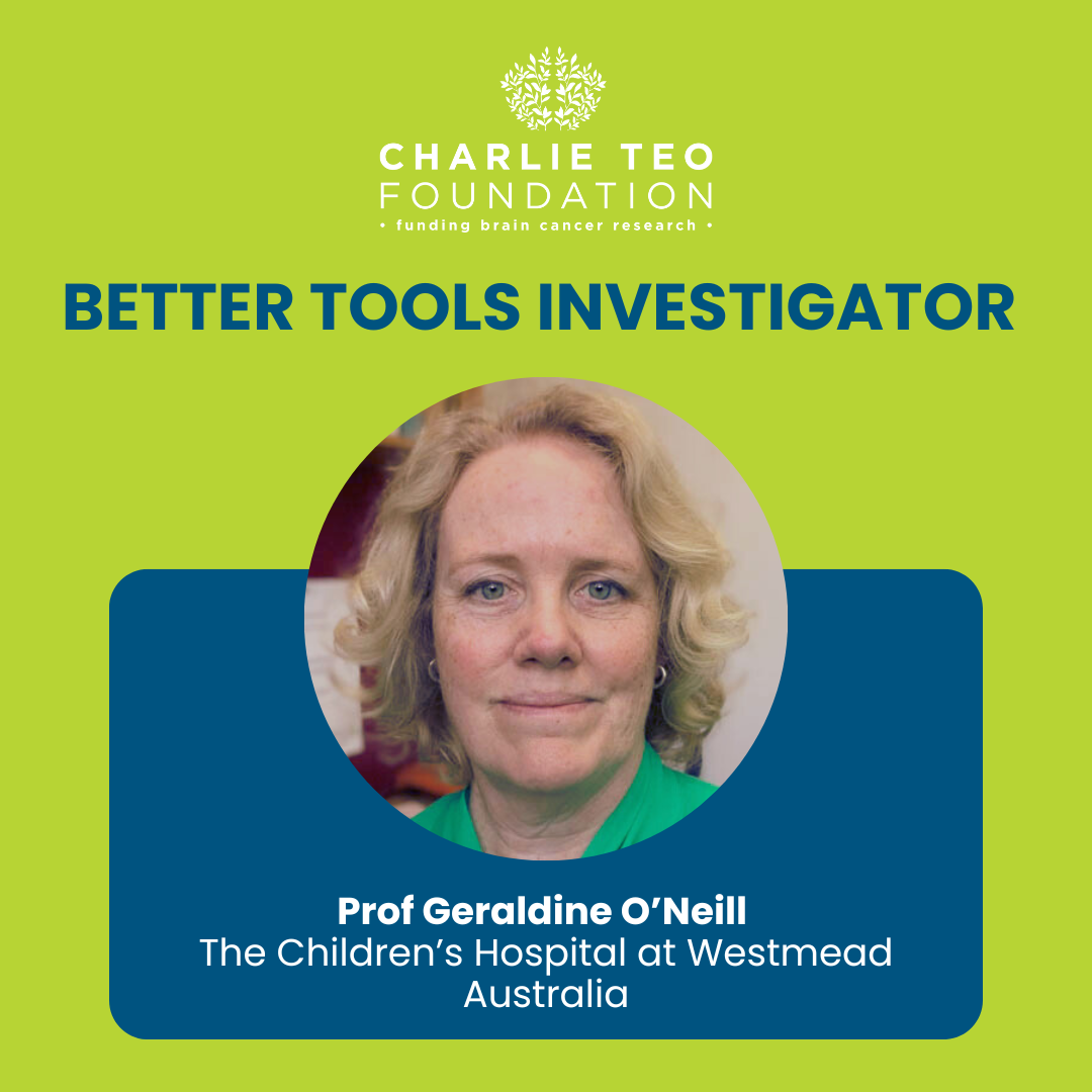
Prof Geraldine O’Neill is Head of the Children’s Cancer Research Unit (CCRU), the research-dedicated arm of the Cancer Centre for Children at the Children’s Hospital at Westmead, and Conjoint Professor with the University of Sydney. Her pioneering research has defined how cancer cells interact with their surrounding tissue environment. Her lab is particularly focused on developing advanced pre-clinical models for brain cancer, leveraging cutting-edge techniques in tissue engineering, cell biology, and biophysics to enhance treatment options for patients. Prof. O’Neill is deeply committed to translating scientific discoveries into new clinical therapies. As the head of the CCRU, her mission is to transform childhood cancer into a thing of the past.
Meet the Researcher
Why is this project game-changing?
This project is game-changing because it uses lab-grown brain structures and patient-derived cancer cells to provide a more accurate assessment of treatment effectiveness for DIPG. Traditional tests often exclude healthy brain cells, which are crucial in understanding treatment responses. This new method has already revealed that some therapies, which seemed promising in standard tests, may not be as effective in real patients. This approach offers a more realistic and rigorous evaluation platform for potential DIPG treatments, potentially leading to better treatment strategies.
How can it potentially help people with brain cancer?
This project can potentially help people with brain cancer by using an improved DIPG model to evaluate new immune cell therapies. By doing so, this will provide a more accurate of how these therapies will perform in real patients. This project aims to broaden the range of available and effective treatments, increasing the chances of finding a cure for every child with brain cancer.
Scientific description
This project aims to develop better preclinical models for pediatric high-grade gliomas (pHGGs), which includes DIPG, by incorporating human stem cell-derived brain organoids and DIPG patient-derived tumouroids. These advanced models will enable more precise testing of new immune cell therapies. The O’Neill lab will utilize this sophisticated DIPG model to evaluate novel CAR T-cell therapies targeting EphA2, which is present in a significant number of pHGG cases, and TCR T-cell therapies targeting PRAME, an antigen over-expressed in various cancers. This project will use liquid biopsies to monitor the effectiveness of these therapies by detecting cell-free DNA, therefore providing a biomarker of intra-tumoural CAR T-cell activity.
The overarching aims in this grant includes:
Aim 1. Assess PRAME TCR T alone and in combination with EphA2 CAR in pHGG. The O’Neill lab aim to address tumour-associated antigen heterogeneity that confounds CAR T treatment of solid tumours and find treatments targeting multiple antigens.
Aim 2. Develop in vitro assays to track T-cell treatment efficacy precisely and accurately in patients. The O’Neill lab’s goal is to develop assays that will use minimally invasive blood/cerebrospinal fluid sampling of cell-free DNA to monitor therapy response and ensure that the observed responses are directly linked to the effectiveness of the CAR T-cell treatment.
Meet the Researcher
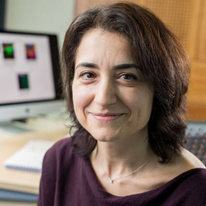
Prof Simona Parrinello is a Professor of Neuro-oncology and Head of the Research Department of Cancer Biology at University College London, UK. She’s also the co-lead of the Cancer Research UK Brain Tumour Centre of Excellence, a joint initiative between UCL and the University of Edinburgh, and is the Samantha Dickson Brain Cancer Unit Lead. Her laboratory investigates the invasive behaviours of glioblastoma tumours with hopes of identifying new therapies for patients.
Why is this project game-changing?
This project is game-changing because studying GBM after standard treatments is extremely difficult due to the rarity and spread of surviving tumour cells throughout the brain. By developing an advanced MRD lab model using patient tumour samples, the Parrinello lab can study these resilient cells without needing risky biopsies. This model will provide a deep understanding of how GBM cells resist treatment and regrow.
How can it potentially help people with brain cancer?
By developing advanced laboratory models that mimic how current treatments for GBM leave behind resilient tumour cells that quickly regrow, this project will enable us to understand the weaknesses of these surviving cells in unprecedented detail. This insight can help create new methods to eliminate these residual cells or make them more responsive to existing treatments, preventing their regrowth into aggressive recurrent tumours. This advanced MRD model will also be used to test new therapies and facilitate personalized drug screening. Ultimately, this could lead to more effective treatments and improved survival rates for GBM patients.
Scientific description
Current standard-of-care treatments for GBM, which include surgical resection, temozolomide chemotherapy, and radiation therapy, fail to prevent rapid tumour recurrence. The persistence of therapy-resistant tumour cells, which invade beyond the resection margin, results in a median survival of less than 15 months. This project aims to develop an ex vivo model of minimal residual disease (MRD) to elucidate the biological vulnerabilities of these residual GBM cells. By leveraging primary and organotypic culture models, mass cytometry, and extensive molecular profiling, this will lead to unprecedented insights into the resistance mechanisms of MRD. This model will serve as a functional test bed for novel therapeutic strategies and facilitate personalized drug screening. The project will also explore the transient cellular states induced by standard-of-care treatment, offering tumour-selective therapeutic opportunities to target these resilient cells to prevent tumour re-growth. Ultimately, this research could lead to more effective, biologically-informed therapies and improved patient outcomes.
The overarching aims of this grant includes:
Aim 1. Develop a model of human GBM residual disease.
Aim 2. Develop a molecular profiling pipeline of resistance mechanisms within residual cells.
Meet the Researcher
Prof. Irina Balyasnikova is a Professor of Neurological Surgery at Northwestern University, Chicago, Illinois. Prof. Balyasnikova completed her Ph.D. at the National Cardiology Research Center, Moscow, Russia, followed by prestigious postdoctoral fellowships at the University of Pennsylvania and the University of Illinois Chicago. She currently leads a research laboratory focused on advancing cell-based therapies for Glioblastoma (GBM) and developing novel imaging approaches for monitoring brain tumours. With over 100 publications and ongoing NIH-supported projects, Prof. Balyasnikova is at the forefront of advancing innovative approaches in brain cancer research.
This project will be carried out in collaboration with experts in molecular and magnetic resonance imaging, Ming Zhao, Ph.D., Associate Professor of Medicine, and Daniele Procissi, Ph.D., Research Professor of Radiology, and the Center for Advance Molecular Imaging (Chad Haney, Ph.D.) of Northwestern University.
Why is this project game-changing?
Prof. Balyasnikova's project is truly game-changing for brain cancer treatment. The development of this novel non-invasive imaging approach is analogous to giving our medical experts a powerful new pair of glasses for the brain, allowing them to witness in real-time how immunotherapy battles GBM. This cutting-edge approach provides a dynamic and detailed view, ensuring quicker and more accurate assessments of treatment effectiveness. By enabling timely adjustments to therapy plans, it not only enhances patient care but also offers the precious gift of time – a crucial factor in the challenging journey against brain cancer. This innovative project represents a significant leap forward, promising a more proactive and effective approach to monitoring GBM and improving outcomes for those facing this formidable disease.
How can it potentially help people with brain cancer?
Prof. Balyasnikova's innovative imaging tool promises clinicians and researchers not only a more reliable but also an earlier indication of therapy effectiveness. This ensures timely adjustments to treatment plans, avoiding unnecessary delays. By incorporating an effective early monitoring program, we can provide patients with the gift of time, enabling a seamless transition to alternative therapies and enhancing the proactive and engaging approach to caring for brain cancer patients.
Scientific description
The recent success of immunotherapy in extracranial malignancies has translated into active clinical trials. However, the standard assessment for therapeutic response relies on conventional CT and MRI, both of which detect primary changes in the tumour (e.g., morphology, size) but fail to reflect the molecular alteration induced by immunotherapy, which often translates into subtle modifications at the cellular level directly linked to positive therapeutic response without early morphological manifestations. In this perspective, clinicians often have difficulty differentiating between actual tumour progression and pseudoprogression (i.e., the complex set of changes that mimic actual progression but, in reality, reflect positive therapeutic outcomes). Developing an imaging approach for monitoring immunotherapy response, which can improve the clinical ability to distinguish between the two regimens, represents an unmet clinical need. The proposed study will employ a preclinical framework aiming to monitor immunotherapeutic response in vivo using a multimodality imaging approach, allowing the detection of spatiotemporal changes in the tumour microenvironment with a particular focus on a quantitative regional assessment of early tumoural apoptosis, which plays an essential role in determining therapeutic outcome early before macroscopic morphological manifestations. The specific strength of our preclinical experimental design is in combined quantitative multiparametric MRI with 99mTc-duramycin SPECT (imaging agent targeting apoptosis) to capture the early changes in cellular and molecular tumour microenvironment linked to immunotherapeutic intervention. This approach will allow the visualization and GBM response to immunotherapy, facilitate toxicity monitoring in the surrounding healthy brain tissue, and identify patient responders and non-responders in the clinic.
Meet the Researcher
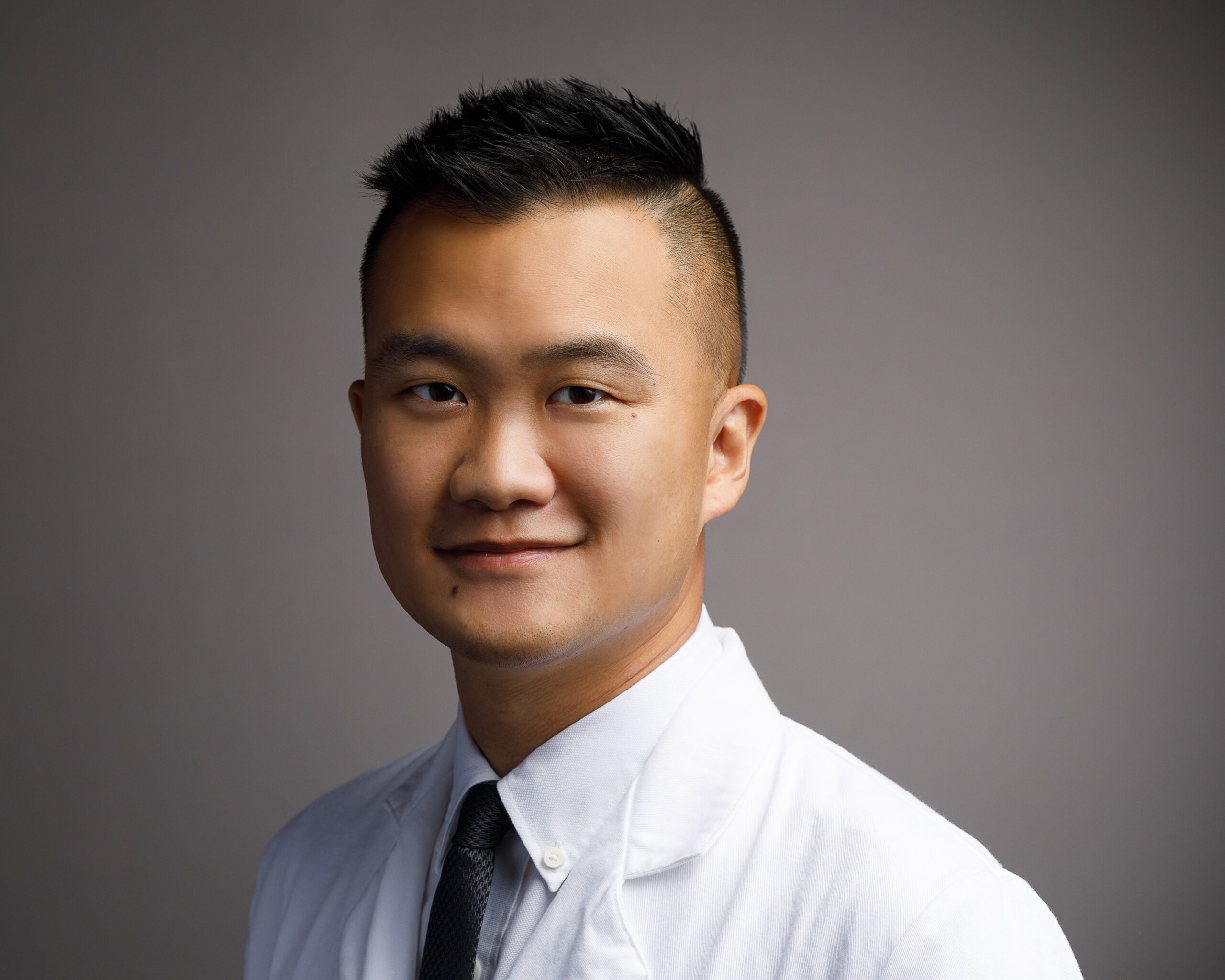
Dr Jacky Yeung is a fellowship-trained neurosurgeon-scientist at Yale University who is an expert on the human brain connectome and studied brain tumour immunology under world-renowned immunologist Dr Lieping Chen. Dr Yeung is among the first to fully characterise the tumour immune microenvironment in malignant meningiomas (a rare brain cancer) and identified a major mechanism by which these tumours evade anti-tumour immunity.
Why is this project game-changing?
Such a molecule has never been discovered before for changing the characteristic of a tumour blood vessel. Its potential for treating brain cancers is simply untapped. We know in real world clinical trials, regular immunotherapy does not work in brain cancers, but the addition of antibodies targeting CD93 may hold the key in unleashing the body's immune system to battle brain cancers with minimal side effects.
How can it potentially help people with brain cancer?
This project aims to resolve one of the greatest problems in tumour immunotherapy, by allowing infiltration of immune cells into the tumour microenvironment, which will enable other scientists to combine their own immunotherapeutic strategies for the treatment of brain cancers. In essence, this treatment strategy is an enabling tool for other immunotherapy researchers to work on further treatments for brain cancer patients.
Scientific description
Targeting CD93/IGFBP7 axis to normalise tumour vasculature and improve T cell trafficking in human gliomas
Tumour vasculature has been theorised to present chemical and physical impedance to effector T cell trafficking. Recently, Dr Yeung’s research group interrogated gene expression profiles in tumours under the treatment of VEGF inhibitors and identified CD93 as a potential target that mediates vascular normalization. The group identified a novel interaction between CD93 and IGFBP7, both overexpressed in tumour but not normal vasculature, that could be antagonised to improve drug delivery and increase immune infiltration.
Aggressive gliomas displayed poor response to nivolumab (anti-PD1 mAb) and overexpression of the IGFBP7/CD93 pathway has been associated with poor response to anti-PD therapies in other cancers. Blockade of the CD93/IGFBP7 using proprietary antibodies developed by the group successfully turned “cold” tumours into “hot” tumours with increased T cell infiltration in preclinical melanoma and pancreatic cancer models.
VEGF overexpression is commonly found in high-grade and recurrent gliomas so CD93-IGFBP7 would likely be induced in these aggressive tumours. Indeed, CD93 expression was recently found to be highly expressed in GBM vasculature, but not in normal brain vessels.
Targeting the CD93/IGFBP7 axis has the potential to enhance the effects of immunotherapy in malignant gliomas without unwanted side-effects of anti-VEGF therapy as CD93/IGFBP7 is downstream in signalling.
Meet the Researcher
A/Prof David Cormode is an Associate Professor of Radiology at University of Pennsylvania, Philadelphia, USA. A/Prof Cormode completed his PhD at University of Oxford, England in the U.K. and is the group leader of the Nanomedicine and Molecular Imaging Lab. In 2020, A/Prof Cormode was awarded the Distinguished Investigator at Academy for Radiology & Biomedical Imaging Research. His research focuses on the development of novel and multifunctional nanoparticle contrast agents for medical imaging applications.
A/Prof Jay Dorsey is an Associate Professor of Radiation Oncology at University of Pennsylvania, USA and Co-Leader of the Radiation Oncology Translational Center of Excellence at the Abramson Cancer Center. A/Prof Dorsey completed his MD and PhD at University of South Florida, USA and is the group leader of the Dorsey Lab. He is also a board-certified neurological radiation oncologist with 14 years’ experience. His research focuses on understanding the underlying mechanisms underpinning cancer resistance to radiation and chemotherapy and characterizing normal cellular responses to radiation therapy.
Why is this project game-changing?
This project combines three game-changing approaches to treating brain cancer: (1) a novel form of radiotherapy – known as FLASH radiotherapy – which uses a rapid, ultra-high dose rate radiotherapy beam. This shortens treatment time and minimises damage to healthy brain tissue (2) injection of a drug-loaded gel (called a hydrogel) into the tumour resection cavity to attack and kill residual cancer cells that could not be surgically removed and (3) the hydrogel loaded with a compound effective at attacking GBM stem cells, the tumour cells responsible for tumour recurrence.
How can it potentially help people with brain cancer?
This project aims to show the effectiveness of drug-loaded gel (known as hydrogel) as a therapy for brain cancer patients, and explore the effectiveness of combining the hydrogel treatment with a rapid, high-dose rate radiotherapy technology. For patients, this combined treatment approach has the potential to minimise treatment time for patients while sparing healthy brain tissue damage. Most importantly, the drug compound in this study has already been shown to effectively kill GBM and glioma stem cells known to drive recurrence, meaning this treatment approach may prevent tumour recurrence.
Scientific description
FLASH radiotherapy and radiation-responsive hydrogel drug delivery as a novel combination therapy for glioblastoma
Glioblastoma (GBM) patients, despite an aggressive treatment strategy, invariably have recurrence of the primary tumour, leading to death. Seeking improved treatments for GBM and the potential glioma stem cells (GSCs) implicated in recurrence, the team performed a high-throughput screen of a large bioactive drug library and found that the drug Quisinostat effectively targets GSCs and GBM cells at nanomolar concentrations. The team also tested this drug and confirmed efficacy in patient-derived GBM organoids and in mouse GBM models, however, the team observed dose-limiting systemic side effects. Therefore, the team now seek to leverage their collective expertise to develop a therapeutic strategy centered on administration of a radiation-responsive drug loaded hydrogel (RR-gel) to the tumour resection cavity. Such an approach will result in high concentrations of drug deposited directly to the target site, while avoiding unnecessary systemic doses to other organs. Furthermore, since radiotherapy is also part of the standard of care, a hydrogel that synergizes with radiation (e.g. increases the effects of radiotherapy and/or releases drug in response to radiotherapy) will improve treatment outcomes.
The team’s promising preliminary data shows that such a hydrogel can effectively control GBM tumours. Moreover, the team plans to integrate the hydrogel with FLASH radiotherapy, a novel form of radiotherapy that involves ultra-fast delivery of radiation treatment at dose rates several orders of magnitude higher than those conventionally used and has the potential to decrease normal tissue toxicity. The team proposes to develop improved versions of our radiation-responsive hydrogel system, characterize them and test them in vitro and in vivo for their anti-GBM efficacy, in combination with FLASH radiotherapy. The team will use spectral CT to monitor the hydrogel in vivo. The safety of the treatment will be extensively assessed. Overall, the team seeks to develop a breakthrough therapy for GBM.


