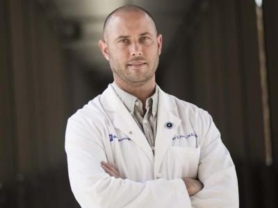
Prof Gelareh Zadeh is a Professor and Dan Chair of Neurosurgery at the University of Toronto, Head of Neurosurgery at Toronto Western Hospital, Co-Director of the Krembil Brain Institute, and Senior Scientist at the Princess Margaret Cancer Research Institute. She was the recipient of the highly prestigious 2023 Canada Gairdner Award for her work on the classification and treatment of brain tumours. The Gairdner is Canada’s most prestigious award for health-related research and approximately a quarter of Gairdner recipients go on to win a Nobel prize.
Meet the Researcher
Why is this project game-changing?
This project is game-changing because it tackles GBM in a novel way using advanced genomics and CRISPR lineage tracing techniques. It focuses on understanding the role of hypoxia, a condition of low oxygen levels within the tumour, and its contribution to the cancer’s aggressiveness and resistance to therapy. The researchers believe that special cells in these low-oxygen areas can survive treatment and cause the cancer to return. By identifying these resistant cells, the project could lead to new targeted therapies that halt tumour progression.
How can it potentially help people with brain cancer?
By understanding the role of hypoxia and cancer stem cells in tumour resistance and recurrence, the project could lead to the development of new therapies that target these aspects of GBM. This could potentially improve treatment outcomes, reduce the likelihood of recurrence, and ultimately, increase survival rates for patients with this aggressive form of cancer.
Scientific Description
This grant is focused on studying the role of hypoxia in GBM recurrence. The team will use spatial transcriptomics and CRISPR/Cas-9 single-cell RNA-seq to understand the genomic and cellular aspects of hypoxia in GBM. They plan to identify hypoxic areas in tumours using pimonidazole, a hypoxia marker, in patients with primary and recurrent GBM as part of their ongoing clinical study. They will compare primary and recurrent patient samples to identify treatment-related alterations in the hypoxic tumour microenvironment. This project aims to understand how hypoxia influences tumour development by studying the lineage of cancer stem cells in hypoxic niches using CRISPR-based in-vivo single-cell lineage tracing xenograft models. The Zadeh lab hypothesize that these cells transition between cellular states to escape therapy mediated by hypoxia. The resulting data will generate a novel atlas of progressive adult gliomas that combines both spatial transcriptomics data and high-quality single-cell clonal information. This will allow them to investigate the regional relationship between cancer cells inside and outside of hypoxic niches, and to track how the cells within these hypoxic niches develop through the use of high-quality phylogenetic trees. The ultimate goal of the research is to uncover key hypoxia-driven signalling pathways that contribute to treatment resistance and GBM recurrence.
The overarching aims of this grant includes:
Aim 1: Decoding the spatial transcriptome architecture of hypoxia in GBM upon recurrence.
Aim 2: Tracing hypoxia-associated tumour cell lineages with CRISPR/Cas-9 Cellular Barcoding.
Meet the Researcher
Prof. Peter Fecci is a neurosurgeon and brain cancer researcher. After completing the MD-PhD program at Duke University, he went on to complete a prestigious residency in neurosurgery at Massachusetts General Hospital and a postdoctoral fellowship at Dana Farber Cancer Institute, Harvard Medical School.
His laboratory’s research focus centres around brain tumour immunology with a specific focus on reversing T-cell dysfunction in patients with glioblastoma and brain metastases. His lab was one of the first to characterize T-cell dysfunction in brain cancer.
Prof Fecci was a member of the Charlie Teo Foundation Scientific Advisory Board (SAB) for over five years from 2018 to 2024. We thank Peter for his immense contribution to the Charlie Teo Foundation and our global scientific impact.
Under our Grant Guidelines and SAB Charter, SAB members must comply with our conflicts of interest policy and relevantly are not involved in peer review of any grant application where they are a principal researcher or member of the research team. Given Prof Fecci’s long-standing service to the SAB, his grant application to the Charlie Teo Foundation was independently reviewed by external reviewers and experts in the field of GBM and cancer immunology.
Why is this project game-changing?
This project is game-changing because it proposes a novel approach to make immunotherapies effective against GBM. By genetically restoring T cell functionality and freeing them from the bone marrow in mice models, the researchers have observed that genetically altered mice can reject GBM and may survive long-term when administered immunotherapies that previously did not work. The project also involves the development of a new class of drug with the help of a Nobel Laureate and a group of medicinal chemists. This drug is anticipated to enhance the success of immunotherapies against GBM by restoring T cell number and function.
How can it potentially help people with brain cancer?
This project can help patients with brain cancer by potentially making immunotherapies effective against GBM. The new class of drug being developed could restore the number and function of T cells, enhancing the body’s immune response against the tumour. This could lead to improved survival rates and quality of life for patients with GBM and potentially other types of cancer that infiltrate the brain.
Scientific Description
This grant is aimed at deriving pharmacologic reversal of T cell sequestration and restoration of T cell function in an effort to license immunotherapeutic success against GBM. The Fecci Lab have previously identified that T cells are sequestered in the bone marrow of patients with intracranial tumours, due to loss of the S1P1 receptor from the T cell surface. They have genetically shown that when S1P1 is restored in mice models, these mice reject GBM and may survive long-term when administered immunotherapies that did not previously work. To translate their findings, they have discovered that a compound that can prevent the loss of S1P1, restoring T- cell function. While this compound works exceedingly well outside the body, it is currently too unstable in the body to be useful as a drug. The Fecci lab has teamed up with a local non-profit group of medicinal chemists from RTI whose expertise is in modifying compounds to make them into useable drugs. This grant proposal aims to take this discovery and invent an entirely new class of drug that they anticipate will newly license immunotherapies success against GBM by restoring T cell number and function.
Meet the Researcher
Professor Matt Dun is a childhood brain cancer researcher focused on finding and/or developing treatments for children with diffuse midline glioma (DMG/DIPG) – the most aggressive childhood brain cancer. Having personally known the hopelessness of hearing that his child’s brain cancer is ‘untreatable’, through his research, Prof Dun is determined to change the ‘go home and make memories’ message that DMG families currently face.
Why is this project game-changing?
Only the genomic features (gene mutations) of a patient’s tumour are analysed at the time of diagnosis due to the limited availability of biopsy material. Although this may identify some genetic characteristics driving tumour growth, few of these mutations are targetable, with even less providing a survival benefit to patients. As DMG tumours are known to be highly heterogeneous (driver mutations vary greatly between and within patient tumours), I-DIMENSIONS seeks to understand the multiple layers influencing DMG across a large selection of tumours so that better-informed treatment strategies can be developed.
How can it potentially help people with brain cancer?
I-DIMENSIONS aims to provide medical and research scientists with new data that unlocks effective treatment strategies for DMG. By looking at tumours as a sum of their biological systems (rather than via a single-featured lens), we hope that the developed subtyping system will inform the creation of a therapy selection tool used by clinicians to extend the survival of children diagnosed with brain cancer in the future.
Scientific Description
I-DIMENSIONS is an Integrated Dmg/hgg genomIc Methylomic EpigeNetic Spatial transcrIptomic prOteomic subtypiNg System integrating tumour genomics data (whole genome sequencing–WGS), DNA methylation data (EPIC array) and chromatin landscapes (ATAC-seq) to group patients into methylo-epi-genomic subtypes at diagnosis. Evaluating the spatial heterogeneity (scRNAseq) and the abundance and activity of all proteins (phospho- and proteomic profiling) present in each specimen will provide the most comprehensive picture of the elements sustaining tumour growth, revealing targets to be addressed using precision medicines. Our approach will be informed by utilising 210 samples collected from collaborators worldwide (including about 17 patient samples from the Charlie Teo Foundation Brain Tumour Bank) and be developed by a multi-disciplinary team of international experts.
Critically, I-DIMENSIONS will identify the highly significant influences on the regulation of a tumour’s posttranslational architecture i.e. the non-genomic elements relating to the geographical location of the tumour. The role that endogenous and exogenous microenvironmental influences (such as neurological cues, catecholamines, insulin, growth factors, growth hormones and immune related interactions - scRNAseq), as well as treatment-related neuronal effects from radiotherapy, and corticosteroid use will dictate posttranscriptional and posttranslational effects, that cannot be replicated in in vitro laboratory models.
Hypothesis:
The development of a methylo-epi-genomic subtyping system predictive of the proteomic/phosphoproteomic components of each sample will enable the future stratification of patients into subtypes that are indicative of treatment response.
Aim 1: Develop a novel methylo-epi-genomic subtyping system for DMG.
Aim 2: Identify the key proteomic/phosphoproteomic signatures (drug targets) of each specimen used in the DMG methylo-epi-genomic subtyping system.
Aim 3: Identify the spatial heterogeneity of the disease and relate this back to the proteomic/phosphoproteomic signatures.
Meet the Researcher
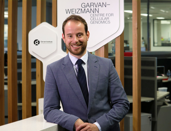
Prof Joseph Powell is a biomedical researcher and geneticist at the Garvan Institute of Medical Research, Australia. Prof Powell completed his PhD at Edinburgh University in 2010 and is now the Director of the Garvan-Weizmann Centre for Cellular Genomics. Prof Powell is a leader in his field, publishing in world-leading journals and having received numerous awards including the NHMRC Research Excellence Medal. He was also the youngest ever recipient of the Commonwealth Health Minister’s Medal and received the Ruth Stephens Gani Medal, an Australian award for outstanding achievement in human genetics.
Why is this project game-changing?
This project will generate a large source of genomics data on brain cancers, a crucial step in understanding the genomic variability of brain cancer. Further, Prof Powell will leverage expertise from machine-learning, biology, genetics and advanced biotechnology domains in an effort to unravel the deep complexities of brain cancer and find effective treatment strategies for brain cancer patients.
How can it potentially help people with brain cancer?
Brain cancer is one of the most lethal cancers, with few effective treatments available for patients. Moreover, a hallmark of brain tumours is its ability to resist treatment due to its complex and variable genetic make-up. By understanding this genetic complexity, Prof Powell’s research team aims to find the ‘bad actors’ responsible for brain cancers resisting treatment and find effective treatments.
Scientific Description
Identifying therapeutic targets from cell state models in gliomas
Glioblastoma is one of the deadliest human cancers, due to its strong resistance to treatment following surgical removal. This resistance is driven by an intrinsic intra-tumoral heterogeneity, which is a hallmark of these tumours. To develop new treatments to overcome this cellular heterogeneity, a more detailed understanding of the nature and drivers of this variation is needed. Single-cell RNA sequencing of human tumours provides a powerful means to systematically interrogate the diversity of malignant and normal cell states.
Recent studies, as well as Prof Powell's work, have highlighted transcriptional cell state diversity across tumour types that is often independent of genetic clonal heterogeneity. His team has developed highly accurate machine learning methods that are able to use the transcriptional signature of a single cell and are able to accurately classify it into a specific cell state. The researchers are able to ‘map’ the within-tumour heterogeneity from thousands of cells in a single patient sample. Doing so over a large clinical cohort across different subtypes of adult diffuse gliomas including astrocytomas, oligodendrogliomas and glioblastomas will allow them to identity the variation in the within-tumour heterogeneity between patients and correlate those cell states with clinical features, recurrence, and treatment resistance. The relationship between cell states and state dynamics will be tested against disease history and clinical features for each cancer classification type (following WHO CNS5 framework).
This project builds on Prof Powell's current research which has been supported by the Charlie Teo Foundation. His team will extend their research into the identification of the genomic drivers of cell states and state dynamics that correlate with clinical outcomes, generating new data on targeted glioblastoma patient cohort, and developing the high-throughput cancer organoid systems to be able to perform high-throughput drug screening against specific tumour cell states.
Meet the Researcher
Prof Peter Fecci is a neurosurgeon, scholar and brain cancer researcher. He has trained at the Ivy League research University, Cornell University, and prestigious Duke University (U.S.). His research centres around brain tumour immunology, immunotherapy and T-cell dysfunction in glioblastoma. He was one of the first to identify T-cell dysfunction in brain cancer.
Why is this project game-changing?
After decades of failure, scientists finally found effective ways of turning the immune system against cancers, with spectacular results – except for brain cancer. Immunotherapies have continued to fail in brain cancer and the team have discovered that the T-cells are stuck in the bone marrow of brain cancer patients. This project may unlock the reason why immunotherapy has failed in brain cancer and lead to immunotherapy treatments finally being effective.
How can it potentially help people with brain cancer?
If the team’s theory is correct, this work will finally allow for the development of effective immunotherapies for patients with brain cancer.
Scientific Description
Development of Beta-arrestin 2 small molecule inhibitors for brain cancer therapy
Cancers of the intracranial (IC) compartment carry unique therapeutic challenges and offer grim outcomes, regardless of tissue type. These cancers include primary malignancies, such as universally lethal glioblastoma (GBM), as well as far more common brain metastases. Our group unveiled the ability of IC tumours to cause a dramatic plunge in the number of circulating T-cells, the main cells that help drive the body’s defences against cancer. Immunotherapy is a novel modality of cancer therapy that has gained momentum in recent years, which works by harnessing and ramping up the body’s own immune system, especially T-cells (the immune system’s effector arm) to fight cancer. However, without T-cells, there is nothing for immunotherapy to act upon. Our group tracked the missing T-cells in patients with IC cancers and discovered them ‘trapped’ in the bone marrow. We showed that this phenomenon is accompanied by loss of sphingosine 1-phosphate receptor 1 (S1P1) from the T-cell surface. S1P1 is a molecule which would normally act as an ‘exit visa’ allowing T-cells to exit from the bone marrow. Without surface S1P1, T-cells instead are locked in, unable to circulate out of the marrow. To solve this problem, our group demonstrated that mice lacking Beta-arrestin 2 (βARR2), an adaptor protein responsible for S1P1 internalisation, do not experience sequestration, and their T-cells are instead able to travel to the brain, and, with the help of immunotherapy, fight their cancers. Our studies in mouse models provided the proof of concept that blocking βARR2 and preventing loss of S1P1 could promote T-cells travelling to the brain to produce anti-tumour responses. However, we’ve now found that inhibiting βARR2 does far more to combat cancer, and we’re finding unprecedented responses against many types of cancer when we knock out βARR2. We think we’ve uncovered an entirely novel cancer therapeutic, and we’ve partnered with a Nobel Laureate who is an expert in this molecule to help develop a pharmacological strategy to block βARR2. We hope that our continued work will allow for the development of more effective immunotherapies for patients with brain cancer.


